Frank - Intestinal Obstruction
Meet Frank! Frank had been boarding at our clinic for a week when he began vomiting. Symptomatic treatment caused the vomiting to cease and Frank did well for several days. He then started having much more profuse vomiting and abdominal pain.
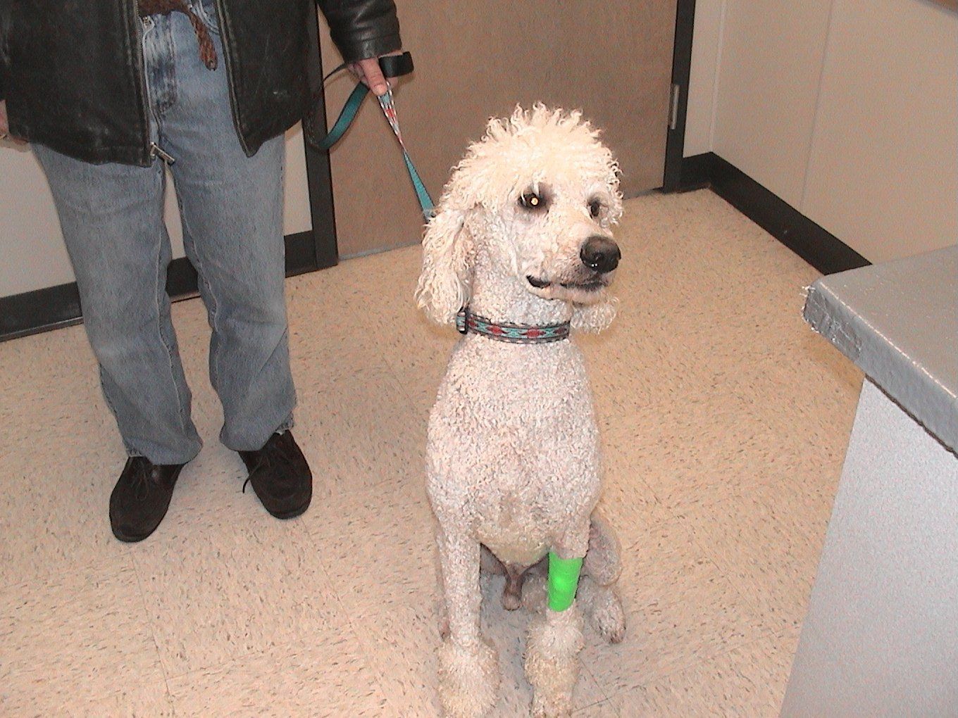
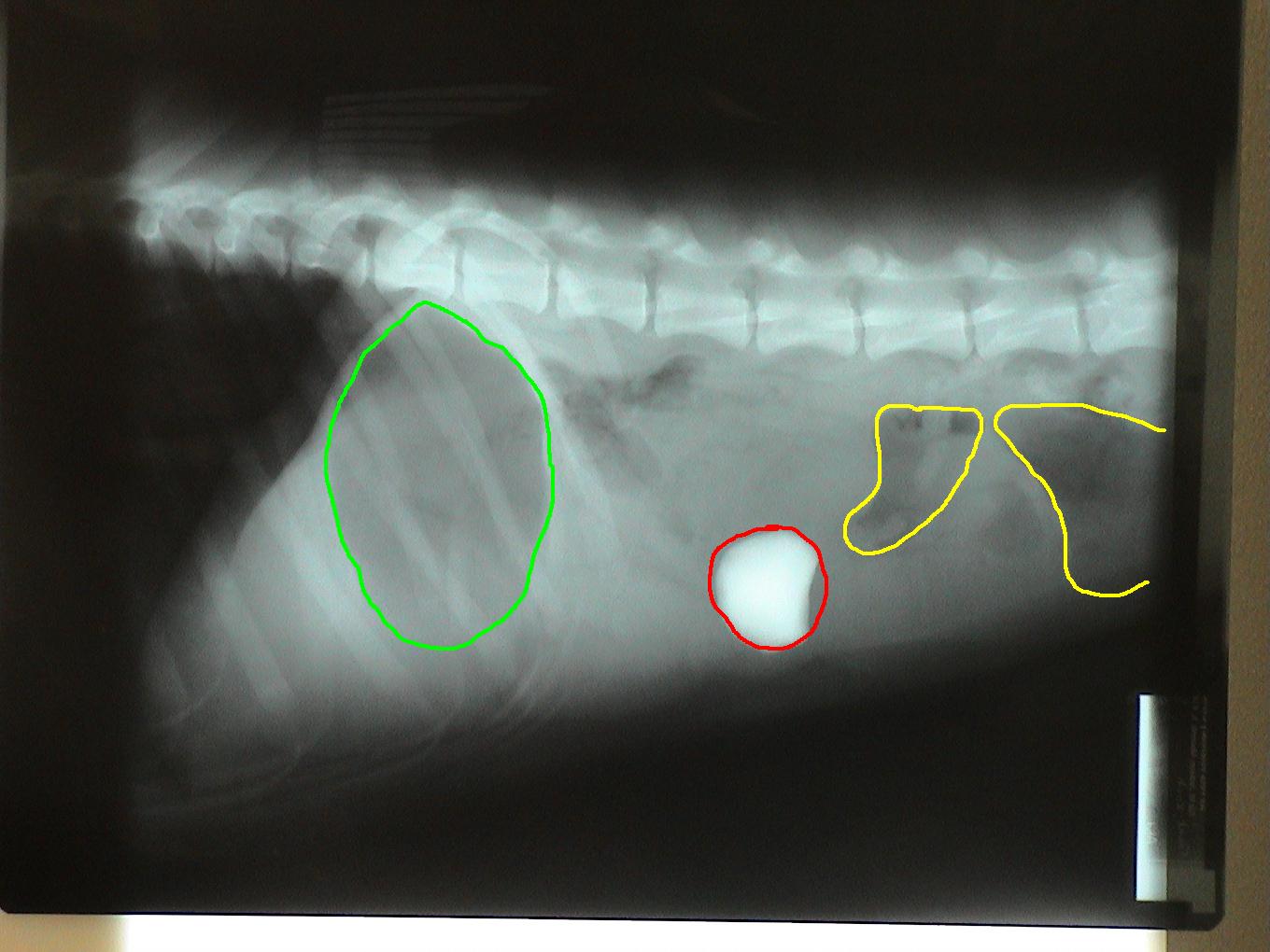
Radiographs (x-rays) were taken and his problem became very obvious! A previously ingested rock had become lodged in the small intestine causing an obstruction.

Green circle: Gas filled stomach. Secondary to vomiting, the stomach can become distended with gas. Red circle: Rock in small intestine. Yellow circle: Gas in colon. Secondary to an obstruction, gas can accumulate in the bowel.
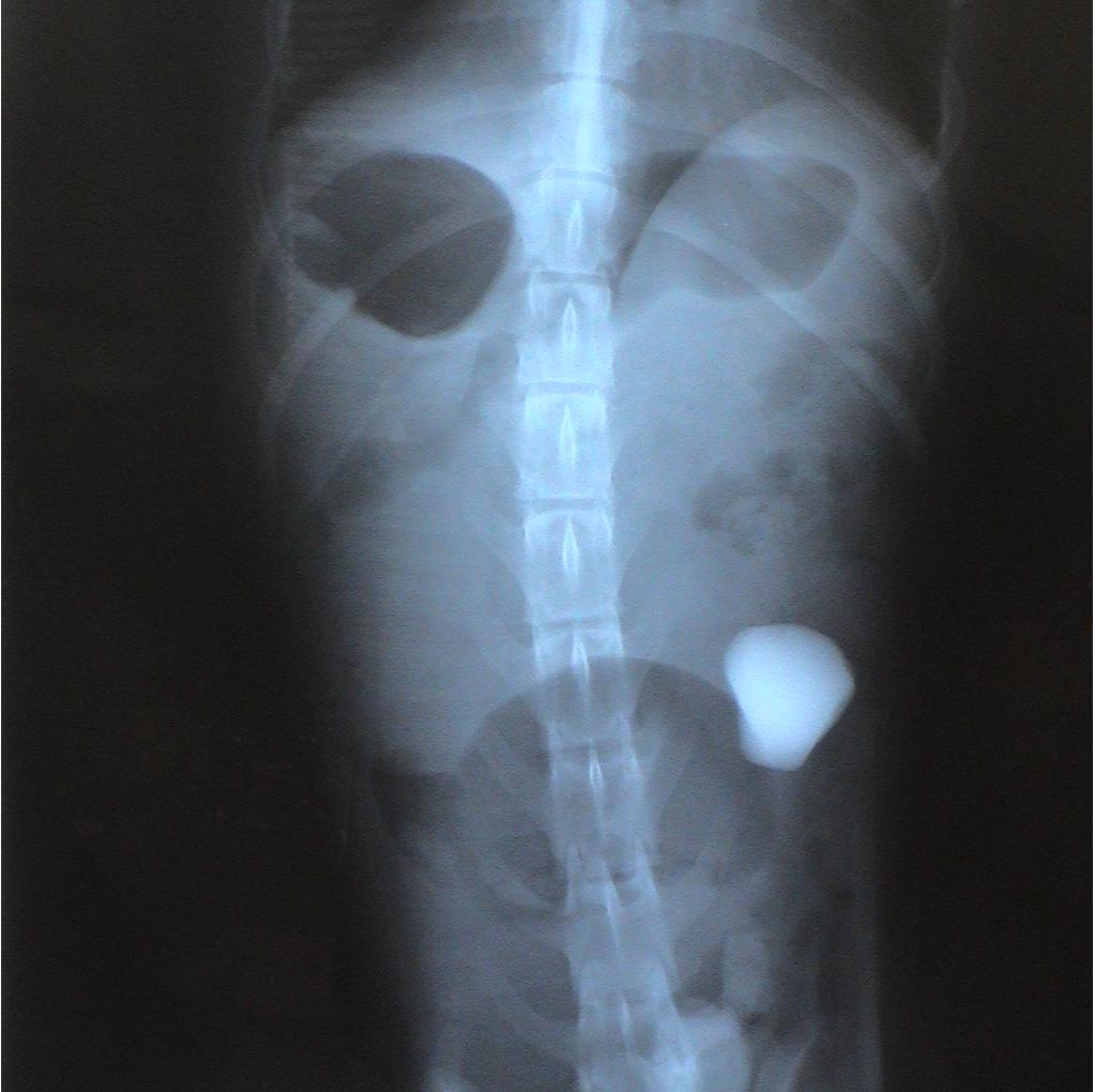
Two sets of radiographs are always taken to localize lesions and identify problems. Radiographs are flat pictures of three-dimensional animals! If one view is taken, things can be lost as structures will overlap each other. For example, the rock is located on the left side of the abdomen which is clearly evident on the second set of radiographs. This is not evident in the first set since the left and right sides of the dog overlap each other.
Notice the difference in the shape of the circled structures depending on the position of the animal.
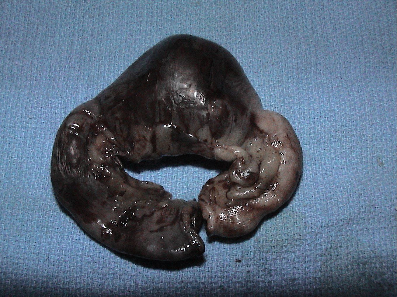
Frank was placed under anesthesia and prepared for surgery. The rock was isolated in the small intestine. Pressure from the rock caused damage to the intestine in the area where the rock had become lodged. Thus, the dying piece of the intestine was removed with the rock in place. This surgery is called an anastomosis and resection.
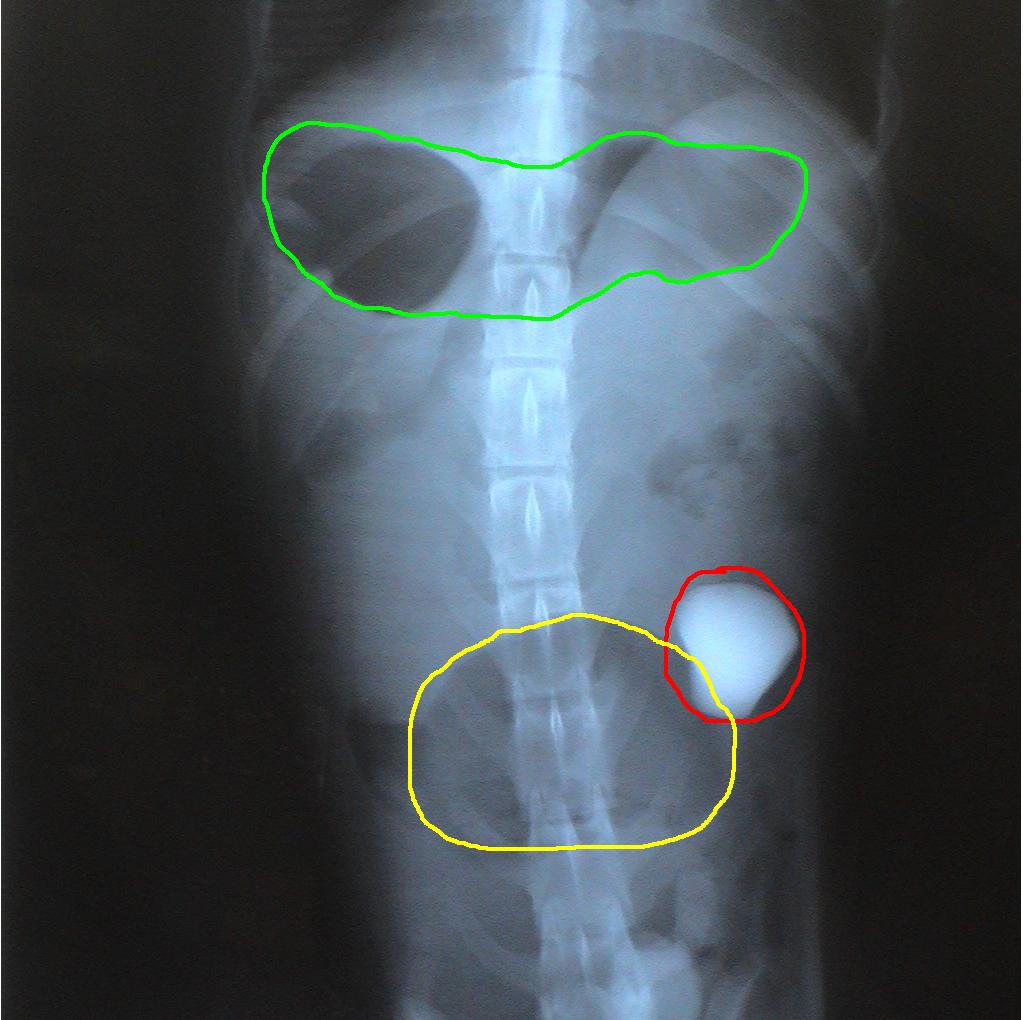
Two sets of radiographs are always taken to localize lesions and identify problems. Radiographs are flat pictures of three-dimensional animals! If one view is taken, things can be lost as structures will overlap each other. For example, the rock is located on the left side of the abdomen which is clearly evident on the second set of radiographs. This is not evident in the first set since the left and right sides of the dog overlap each other.
Notice the difference in the shape of the circled structures depending on the position of the animal.
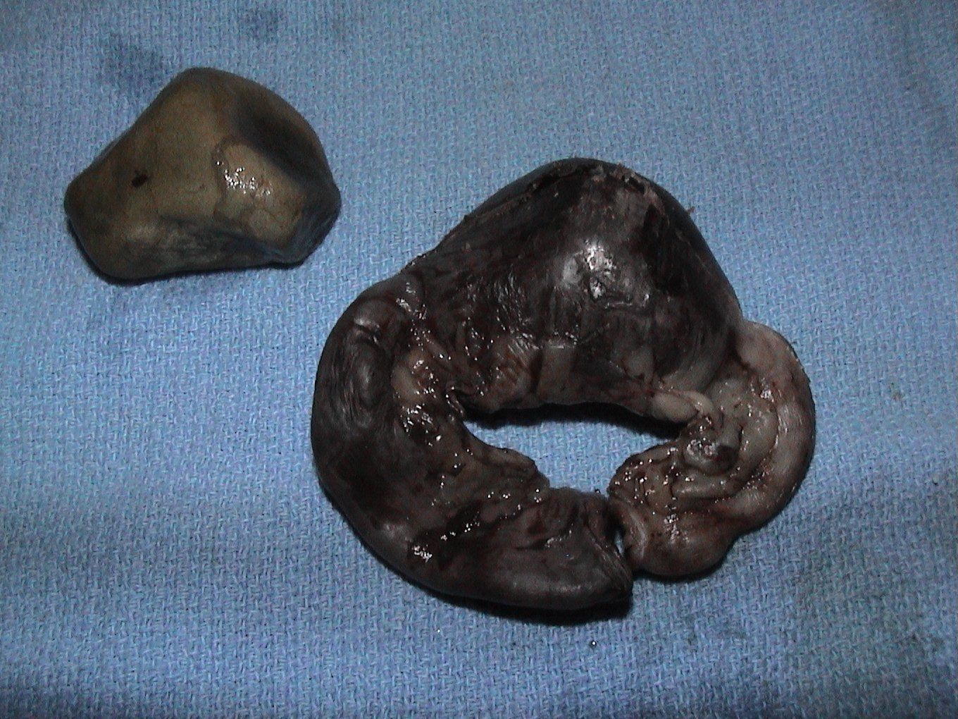
In this picture (below), the rock has been removed from the piece of resected intestine.
Frank recovered fully after surgery and is doing very well. The owner noted that Frank had been having sporadic episodes of vomiting over the previous 4-5 weeks. We are assuming that the rock was present in his stomach for that entire duration of time. The rock did not cause serious problems until it finally passed out of the stomach and lodged in a small area of the small intestine.
Special thanks to Frank's owner for allowing us to share this case.
Learn More About
All Pets Veterinary Clinic

Share On: